WHAT ARE MEDICAL
X-RAYS?
X-rays, a form of electromagnetic radiation similar to visible light, are used in a safe and controlled manner. Unlike light, however, X-rays have higher energy and can pass through most objects, including the body.
Medical X-rays are used to generate highly detailed images of tissues and structures inside the body. If X-rays traveling through the body also pass through an X-ray detector on the other side of the patient, an image will be formed that represents the “shadows” formed by the objects inside the body.
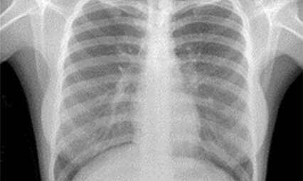
One type of X-ray detector is photographic film; however, there are many other types of detectors used to produce digital images. The X-ray images that result from this process, known as radiographs, are essentially photographs of the internal structures of the body, captured using X-rays instead of visible light.
 Request An Appointment
WHAT ARE DIGITAL
X-RAYS?
A digital X-ray, also known as digital radiography, is a modern type of X-ray that utilizes digital sensors instead of photographic film, similar to traditional X-rays. The image captured is converted to digital data immediately, saving valuable time, and is available for review within seconds.
This enables the modern X-ray to capture images with an unprecedented level of precision, providing healthcare professionals with a high degree of accuracy in their diagnoses.
HOW IT WORKS
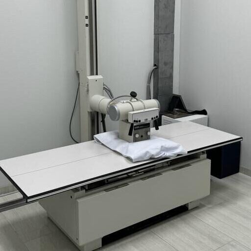
The creation of a radiograph is a sophisticated process that involves the precise positioning of a patient. This positioning ensures that the part of the body being imaged is accurately located between an X-ray source and an X-ray detector. When the machine is turned on, X-rays, the key players in this process, travel through the body and are absorbed in varying amounts by different tissues, depending on the radiological density of the tissues they pass through.
Radiological density, a key concept in radiography, is a measure of how much a material resists the passage of X-rays. It is determined by both the density and the atomic number (the number of protons in an atom’s nucleus) of the materials being imaged. For example, structures such as bone contain calcium, which has a higher atomic number than most tissues. Because of this property, bones readily absorb X-rays and, thus, produce high contrast on the X-ray detector.
As a result, bony structures stand out distinctly, appearing whiter than other tissues against the black background of a radiograph. This stark contrast is a key feature of radiographs. Conversely, X-rays travel more easily through less radiologically dense tissues, such as fat and muscle, as well as through air-filled cavities, like the lungs. These structures are displayed in shades of gray on a radiograph, creating a nuanced visual representation of the body’s internal structures.
Get a Digital X-Ray Scan at Optimum Imaging TODAY!
X-ray Frequently Asked Questions
What happens during an X-ray?
X-rays are fast, easy, and painless. Once the X-ray machine is carefully aimed at the part of the body being examined, it produces a small burst of radiation that passes through the body, recording an image digitally.
How should I prepare for an X-ray?
We recommend wearing comfortable, athletic-style clothing. If your clothing has metal or other objects in or on it (such as zippers or buttons), you may be asked to change into a gown to minimize the interference caused by these metal objects with the imaging technique.
Are X-rays safe?
When you get an X-ray, a part of your body is exposed to a small dose of ionizing radiation to create images of the inside. X-rays, a form of radiation similar to light or radio waves, can pass through most objects, including the human body. It’s important to remember that X-rays are safe when used by radiologists and technologists who are specially trained to minimize exposure. Their expertise ensures that no radiation remains in your body after the X-ray is taken, reinforcing the safety of the procedure.
Choose accurate imaging and convenient service with Optimum Imaging
At Optimum Imaging Center, we stand out with our unwavering commitment to accurate and timely diagnostic imaging. Our dedication to patient care, use of cutting-edge technology, and focus on your well-being set us apart in the healthcare industry.
When you trust us with your imaging needs, you’re not just another patient. You’re a priority. Experience the personalized care and attention that sets us apart in the industry. We’re here to serve you and your health needs.


